Step into the world of advanced surgical procedures at Levantine Dental Clinic in Dubai. Our Oral & Maxillofacial Surgery offers a comprehensive, precise, and expert solution for complex dental issues. Conducted by skilled surgeons, this surgery is your pathway to improved oral health and function.
BOOK AN APPOINTMENTWhat is Oral and Maxillofacial Surgery? When do patients need to undergo maxillofacial surgery? The specialty of oral and maxillofacial surgery is a surgical discipline of dentistry that encompasses the art and science of diagnosing and treating the diseases, injuries, and defects of the hard and soft tissues of the maxillofacial region and oral cavity. An oral surgical practice deals with the initial and definitive care of the trauma patient, and management of extensive infection of the head and neck. Others include management of pathological conditions such as cysts and tumors, diagnosis and management of dentofacial deformities, complex maxillofacial pre-prosthetic surgery, reconstruction with bone grafts, and management of facial pain and Temporomandibular Joint (TMJ) disorders.
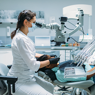
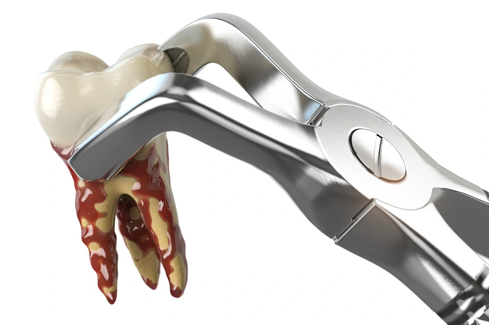
Wisdom teeth, or third molars, typically emerge in late adolescence or early adulthood and can cause problems for many patients. To prevent future dental issues, it is recommended that wisdom teeth be removed at a younger age when the teeth are not fully formed, making surgery easier. When wisdom teeth do emerge, they may be difficult to keep clean and can impact the teeth in front of them, leading to further problems. In some cases, the teeth may be impacted underneath the gums, which can cause cysts and other complications. The removal process typically takes 45 to 60 minutes and is done under sedation. The complexity of the surgery depends on various factors, such as the position of the tooth, the curvature of the roots, and the thickness of the surrounding bone. After the surgery, patients are given instructions and prescriptions before leaving to rest and recover at home.
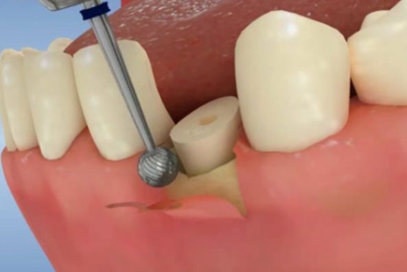
Sometimes, a cavity can develop underneath the gums and cannot be accessed for restoration without cutting part of the gum and reducing bone height. The dentist can then access the cavity and restore it with a resin filling. If a cusp breaks off a tooth below the gums, the tooth cannot be restored by the dentist alone. However, a surgeon can perform a crown lengthening procedure by raising a flap of gum tissue, reducing the bone level, and suturing the flap back in place. After healing, the dentist can restore the tooth using a resin filling or crown. This procedure is called crown lengthening because it makes the crown appear longer.
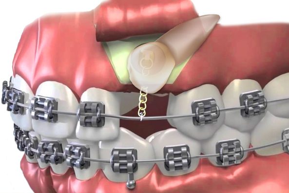
When teeth are trapped underneath the gums, or grow partially into the mouth, this is called impaction. An impacted tooth means that it is stuck and cannot erupt into function. This occurs when there is not enough space in the mouth for teeth to emerge properly. Some impacted teeth can simply be extracted. However, impacted canines and other adult teeth must be guided into their proper place for a healthy and functional bite. At Levantine Dental Clinic, the state-of-the-art CBCT 3D X-ray technology is critical in helping diagnose impacted teeth and pinpoint their exact location. First, the dentist uses braces to make room for the impacted tooth. The patient’s oral surgeon will carefully expose the tooth by removing the overlying gum and bone. A very small bracket and chain are then attached to the tooth. The special loop wire puts tension on the chain, slowly guiding the impacted tooth into place with the braces. Treating impacted teeth early is essential to creating a stable bite and avoiding costly orthodontic or restorative work later in life. At Levantine Dental Clinic, we focus on our patients’ needs to provide them with a healthy lifelong smile.
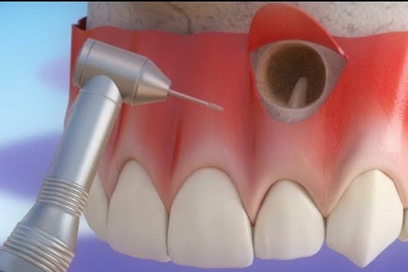
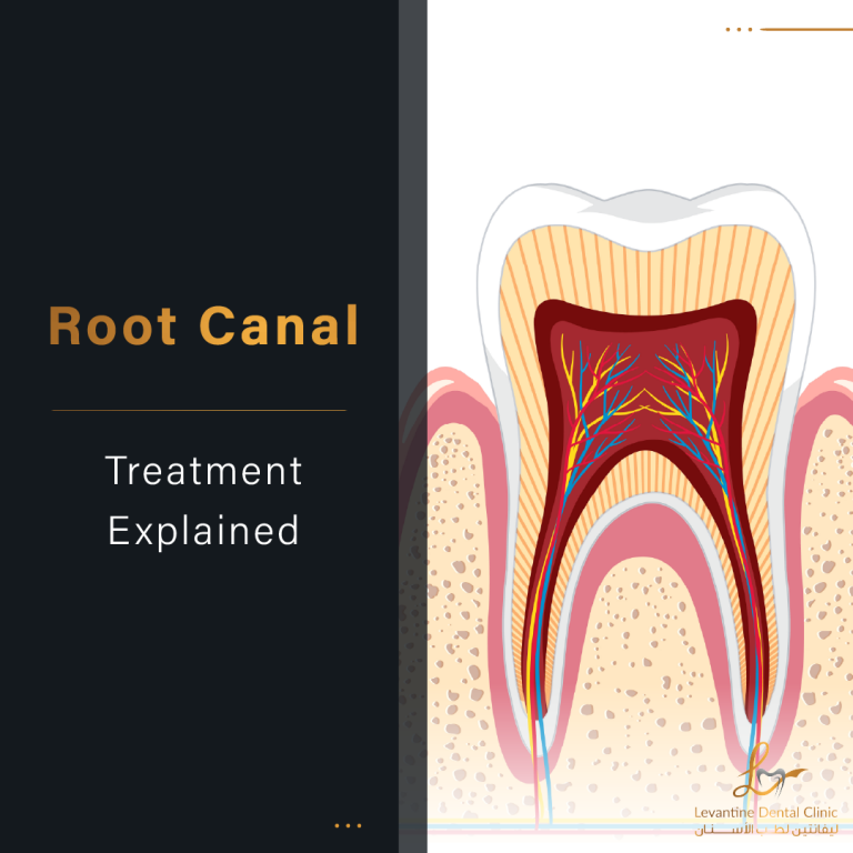
An apicoectomy is a simple surgical procedure performed on an infected tooth under local anesthesia. It involves the removal of the apex, or root tip, of the tooth to address a persistent infection. Unlike a root canal, which treats inflammation and infection in the inner pulp tissue of the tooth, an apicoectomy addresses the infection affecting the last 3mm of the root. The procedure is usually done after a root canal treatment to fix the root or tissues around the tooth. During the procedure, the root tip is sectioned under magnification with a microscope, and a small incision is made in the gum to access the area of infection. The root tip is then addressed and filled. An apicoectomy is typically used to save a tooth that has already had a root canal but has become reinfected. Other reasons for the procedure include treating a fracture or a root with a hole, and addressing bone loss that causes loose teeth. The procedure is painless and done under anesthesia.
In some cases, the dentist removes a tooth without placing a dental implant during the same visit. The jawbone naturally shrinks when there is no tooth, leading to great bone loss. To have a dental implant later during another visit, the patient needs more bone to place the implant. Different bone-grafting techniques trigger bone regeneration to allow the patient's jaw to accept the implant.
This is done during the patient's visit for a tooth removal. After the surgeon removes the tooth, the surgeon places grafting material within the hole. Then, bone healing takes about 3 or 4 months. After 4 months, the surgeon carries out the dental implant procedure.
This technique is for patients who need more bone to carry out a dental implant procedure. The surgeon places blocks of bone onto the space where the bone is missing. The surgeon places the blocks of bone with screws. After 5 months, when the bone heals, the surgeon can carry out the dental implant procedure.
This technique is for places where the surgeon cannot use blocks of bone. The surgeon places the substitutes where the bone is missing and covers the area with a membrane. After a while when the bone has healed, the surgeon can carry out the dental implant.
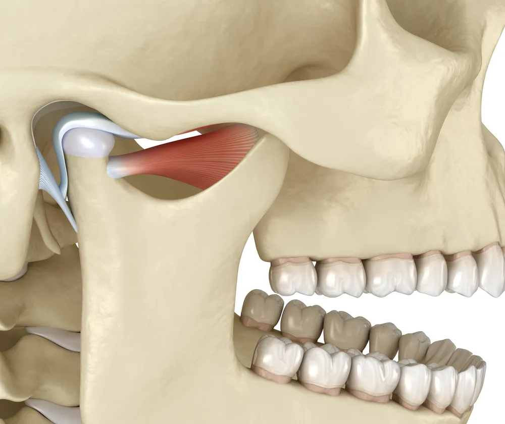
The temporomandibular joint (TMJ) is where the lower jaw and base of the skull meet, forward of the ears. The TMJ is a compound joint involving 3 bones in the body. The mandible, the temporal bone, and the articular disk. We put an amount of pressure on this joint as we breathe, speak, and chew. As we open our mouths the movement begins and as we continue to open our mouths more, the lower jaw starts to slide downward and forward.
Patients notice TMD by experiencing pain in the temporomandibular joint or the muscles around it. The disk is a component of the temporomandibular joint and it acts as a pillow absorbing the shock to protect the joint. When the disk remains in its correct position while opening and closing the jaw, no clicking or popping should occur. However, if there is conflict in the joint, it makes popping sounds upon opening and closing the jaw.
Several conditions that occur in TMD situations.
Myofascial Pain is the most popular temporomandibular joint disorder. It causes pain in the jaw muscles and the tissue around them, as well as the neck and shoulder.
TMJ Internal Derangement is the dislocation of the jaw or the disk that acts as a pillow or shock absorber between the head of the jawbone and the skull. This TMD could also be caused by an injury to the condyle, the rounded end of the jawbone that forms the articulation with the temporal skull bone.
Degenerative Joint Disease is caused by osteoarthritis (when the protective cartilage that cushions the ends of the bones wears down) or rheumatoid arthritis (an autoimmune and inflammatory disease) in the jaw joint.
The treatment of temporomandibular joint disorder depends on how severe each case is. It also depends on the age of the patients and their overall health.
It consists of placing removable dental appliances that provide protection for the temporomandibular joint and helps keep it in place. This reduces inflammation and pain. However, this is merely a treatment, it is not a cure. It will not restore any damage.
It is similar to splint therapy, however, an orthotic is an acrylic device that the patients wear on their lower teeth to improve the function of the temporomandibular joint. This decompresses the joint, which reduces inflammation and helps restore the proper muscle length. Surgery is recommended in very few TMD cases.
Extraction is recommended when wisdom teeth have problems such as pain, infection or decay, or when they might cause damage to other teeth or lead to crowding. Your dentist will evaluate your specific situation with X-rays to determine the best course of action.
Your dentist will administer local anesthetic to numb the area. If the tooth is impacted, the dentist will cut away gum and bone tissue that cover the tooth, then grasp the tooth with forceps and gently loosen it. The procedure is comfortable and recovery is typically straightforward.
Diagnostic imaging includes routine dental X-rays, CT/CAT scans for detailed bone structure analysis, MRI for disc and soft tissue visualization, and bone scans (scintigraphy) for comprehensive joint assessment. A thorough clinical examination is also performed.
Disclose your complete medical history, all medications you are taking, and any conditions including damaged heart valves, congenital heart defects, impaired immunity, liver disease, artificial joints, or history of bacterial endocarditis. This information is crucial for safe treatment planning.
Avoid rinsing for 12 hours after the procedure. Keep the area clean, and avoid eating or drinking until anesthesia wears off. Manage bleeding with gentle pressure using gauze. Take prescribed painkillers within 24 hours post-op. Avoid smoking for at least 5 days after the extraction.
Bone grafting is a surgical procedure that replaces missing bone in order to gain bone volume, thickness and length. This is commonly needed before dental implant placement when the jawbone has deteriorated due to tooth loss or other conditions.